ตำรายาของประเทศไทย
Thai Pharmacopoeia
สำนักยาและวัตถุเสพติด กรมวิทยาศาสตร์การแพทย์ กระทรวงสาธารณสุข
Bureau of Drug and Narcotic, Department of Medical Sciences, Ministry of Public Healthสำนักยาและวัตถุเสพติด กรมวิทยาศาสตร์การแพทย์ กระทรวงสาธารณสุข
Bureau of Drug and Narcotic, Department of Medical Sciences, Ministry of Public Health4.27 PARTICULATE MATTER IN INJECTIONS
Particulate Contamination: Subvisible Particles
Particulate matter consists of mobile, randomlysourced, extraneous substances, other than gas bubbles, that cannot be quantitated by chemical analysis due to the small amount of material that it represents and to its heterogeneous composition. Injectable solutions, including solutions constituted from sterile solids intended for parenteral use, are essentially free from particulate matter that can be observed on visual inspection. The tests described herein are physical tests performed for the purpose of enumerating subvisible extraneous particles within specific size ranges.
Microscopic and light obscuration procedures for the determination of particulate matter are given herein. This appendix provides a test approach in two stages. The injection is first tested by the light obscuration procedure (stage 1). If it fails to meet the prescribed limits, it must pass the microscopic procedure (stage 2) with its own set of test limits. Where for technical reasons the injection cannot be tested by light obscuration, microscopic testing may be used exclusively. Documentation demonstrating that the light obscuration procedure is incapable of testing the injection or produces invalid results is required in each case. It is expected that most articles will meet the requirements on the basis of the light obscuration test alone; however, it may be necessary to test some articles by the light obscuration test followed by the microscopic test to reach a conclusion on conformance to requirements.
All large-volume injections for single-dose infusion and those small-volume injections for which the monographs specify such requirements are subject to the particulate matter limits set forth for the test being applied, unless otherwise specified in the individual monograph. Excluded from the requirements of this appendix are injections intended solely for intramuscular and subcutaneous administration.
Not all injection formulations can be examined for particles by one or both of these tests. Any product that is not a pure solution having a clarity and a viscosity approximating those of water may provide erroneous data when analyzed by the light obscuration counting method. Such materials may be analyzed by the microscopic method. Emulsions, colloids and liposomal preparations are examples. Similarly, products that produce air or gas bubbles when drawn into the sensor, such as bicarbonate-buffered formulations, may also require microscopic testing. Refer to the specific monographs when a question of test applicability occurs. Higher limits are appropriate for certain articles and will be specified in the individual monographs.
In some instances, the viscosity of a material to be tested may be sufficiently high so as to preclude its analysis by either test method. In this event, a quantitative dilution with an appropriate diluent may be made to decrease viscosity, as necessary, to allow the analysis to be performed.
In the tests described below for large-volume and small-volume injections, the results obtained in examining a discrete unit or group of units for particulate matter cannot be extrapolated with certainty to other units that remain untested. Thus, statistically sound sampling plans based upon known operational factors must be developed if valid inferences are to be drawn from observed data to characterize the level of particulate matter in a large group of units. Sampling plans should be based on consideration of product volume, numbers of particles historically found to be present in comparison to limits, particle size distribution of particles present, and variability of particle counts between units.
Light obscuration particle count test Use a suitable apparatus based on the principle of light blockage which allows an automatic determination of the size of particles and the number of particles according to size.
The apparatus is calibrated using suitable certified reference materials consisting of dispersions of spherical particles of known sizes between 10 μm and 25 μm. These standard particles are dispersed in particle-free water. Care must be taken to avoid aggregation of particles during dispersion.
GENERAL PRECAUTIONS The test is carried out under conditions limiting particulate contamination, preferably in a laminar-flow cabinet.
Very carefully wash the glassware and filtration equipment used, except for the membrane filters, with a warm detergent solution and rinse with abundant amounts of water to remove all traces of detergent. Immediately before use, rinse the equipment from top to bottom, outside and then inside, with particle-free water.
Take care not to introduce air bubbles into the preparation to be examined, especially when fractions of the preparation are being transferred to the container in which the determination is to be carried out.
In order to check that the environment is suitable for the test, that the glassware is properly cleaned and that the water to be used is particle-free, the following test is carried out: determine the particulate contamination of five samples of particle-free water, each of 5 ml, according to the procedure described below. If the number of particles of 10 μm or more size exceeds 25 for the combined 25 ml, the precautions taken for the test are not sufficient.
The preparatory steps must be repeated until the environment, glassware and water are suitable for the test.
PROCEDURE Mix the contents of the sample by slowly inverting the container 20 times successively. If necessary, cautiously remove the sealing closure. Clean the outer surfaces of the container opening using a jet of particle-free water and remove the closure, avoiding any contamination of the contents. Eliminate gas bubbles by appropriate measures such as allowing to stand for 2 minutes or sonicating.
For large-volume parenterals, single units are tested. For small-volume parenterals less than 25 ml in volume, the contents of 10 or more units are combined in a cleaned container to obtain a volume of not less than 25 ml; where justified and authorized, the test solution may be prepared by mixing the contents of a suitable number of vials and diluting to 25 ml with particle-free water or with an appropriate solvent without contamination of particles when particle-free water is not suitable. Small-volume parenterals having a volume of 25 ml or more may be tested individually.
Powders for parenteral use are reconstituted with particle-free water or with an appropriate solvent without contamination of particles when particle-free water is not suitable.
The number of test specimens must be adequate to provide a statistically sound assessment. For largevolume parenterals or for small-volume parenterals having a volume of 25 ml or more, less than 10 units may be tested, based on an appropriate sampling plan.
Remove 3 portions, each of not less than 5 ml, and count the number of particles equal to or more than 10 μm and 25 μm. Disregard the result obtained for the first portion, and calculate the mean number of particles for the preparation to be examined.
INTERPRETATION The preparation meets the requirements of the test if the calculated number of particles present in each discrete unit tested or in each pooled sample tested does not exceed the appropriate value listed in Table 1. If the average number of particles exceeds the limit, test the article by Microscopic Particle Count Test.
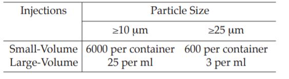
Table 1 Maximum Limit of Number of Particles by Light Obscuration Test
Microscopic particle count test Use a suitable binocular microscope, filter assembly for retaining particulate contamination and membrane filter for examination.
The microscope is equipped with an ocular micrometer calibrated with an objective micrometer, a mechanical stage capable of holding and traversing the entire filtration area of the membrane filter, two suitable illuminators to provide episcopic illumination in addition to oblique illumination, and is adjusted to 100±10 magnifications.
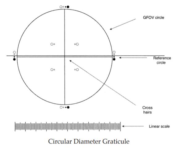
The ocular micrometer is a circular diameter graticule (see Figure) and consists of a large circle divided by crosshairs into quadrants, transparent and black reference circles 10 μm and 25 μm in diameter at 100 magnifications, and a linear scale graduated in 10 μm increments. It is calibrated using a stage micrometer that is certified by either a domestic or international standard institution. A relative error of the linear scale of the graticule within ±2 per cent is acceptable. The large circle is designated the graticule field of view (GFOV).
Two illuminators are required. One is an episcopic brightfield illuminator internal to the microscope, and the other is an external, focusable auxiliary illuminator adjustable to give reflected oblique illumination at an angle of 10º to 20º.
The filter assembly for retaining particulate contamination consists of a filter holder made of glass or other suitable material, and is equipped with a vacuum source and a suitable membrane filter.
The membrane filter is of suitable size, black or dark grey in colour, non-gridded or gridded, and 1.0 μm or finer in nominal pore size.
GENERAL PRECAUTIONS The test is carried out under conditions limiting particulate contamination, preferably in a laminar-flow cabinet.
Very carefully wash the glassware and filter assembly used, except for the membrane filter, with a warm detergent solution and rinse with abundant amounts of water to remove all traces of detergent. Immediately before use, rinse both sides of the membrane filter and the equipment from top to bottom, outside and then inside, with particle-free water.
In order to check that the environment is suitable for the test, that the glassware and the membrane filter are properly cleaned and that the water to be used is particle-free, the following test is carried out: determine the particulate contamination of a 50 ml volume of particle-free water according to the procedure described below. If more than 20 particles 10 μm or larger in size or if more than 5 particles 25 μm or larger in size are present within the filtration area, the precautions taken for the test are not sufficient.
The preparatory steps must be repeated until the environment, glassware, membrane filter and water are suitable for the test.
PROCEDURE Mix the contents of the samples by slowly inverting the container 20 times successively. If necessary, cautiously remove the sealing closure. Clean the outer surfaces of the container opening using a jet of particle-free water and remove the closure, avoiding any contamination of the contents.
For large-volume parenterals, single units are tested. For small-volume parenterals less than 25 ml in volume, the contents of 10 or more units are combined in a cleaned container; where justified and authorized, the test solution may be prepared by mixing the contents of a suitable number of vials and diluting to 25 ml with particle-free water or with an appropriate solvent without contamination of particles when particle-free water is not suitable. Small-volume parenterals having a volume of 25 ml or more may be tested individually. Powders for parenteral use are constituted with particle-free water or with an appropriate solvent without contamination of particles when particle-free water is not suitable.
The number of test specimens must be adequate to provide a statistically sound assessment. For largevolume parenterals or for small-volume parenterals having a volume of 25 ml or more, less than 10 units may be tested, based on a appropriate sampling plan.
Wet the inside of the filter holder fitted with the membrane filter with several millilitres of particle-free water. Transfer to the filtration funnel the total volume of a solution pool or of a single unit, and apply vacuum. If needed, add stepwise a portion of the solution until the entire volume is filtered. After the last addition of solution, begin rinsing the inner walls of the filter holder by using a jet of particle-free water. Maintain the vacuum until the surface of the membrane filter is free from liquid. Place the filter in a Petri dish and allow the filter to air-dry with the cover slightly ajar. After the filter has been dried, place the Petri dish on the stage of the microscope, scan the entire membrane filter under the reflected light from the illuminating device, and count the number of particles that are equal to or greater than 10 μm and the number of particles that are equal to or greater than 25 μm. Alternatively, partial filter count and determination of the total filter count by calculation is allowed. Calculate the mean number of particles for the preparation to be examined.
The particle sizing process with the use of the circular diameter graticule is carried out by transforming mentally the image of each particle into a circle and then comparing it to the 10 μm and 25 μm graticule reference circles. Thereby the particles are not moved from their initial locations within the graticule field of view and are not superimposed on the reference circles for comparison. The inner diameter of the transparent graticule reference circles is used to size white and transparent particles, while dark particles are sized by using the outer diameter of the black opaque graticule reference circles.
In performing the microscopic particle count test do not attempt to size or enumerate amorphous, semiliquid, or otherwise morphologically indistinct materials that have the appearance of a stain or discoloration on the membrane filter. These materials show little or no surface relief and present a gelatinous or film-like appearance. In such cases the interpretation of enumeration may be aided by testing a sample of the solution by the light obscuration particle count test.
INTERPRETATION The preparations meets the requirements of the test if the number of particles present (actual or calculated) in each discrete unit tested or in each pooled sample tested does not exceed the appropriate value listed in Table 2.
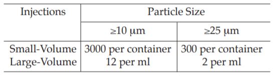
Table 2 Maximum Limit of Number of Particles by Microscopic Particle Count Test
Particulate Contamination: Visible Particles
Particulate contamination of injections and infusions consists of extraneous, mobile undissolved particles, other than gas bubbles, unintentionally present in the solutions. The test is intended to provide a simple procedure for the visual assessment of the quality of parenteral solutions as regards visible particles. Other validated methods may be used.
APPARATUS The apparatus consists of a viewing station comprising: a matt black panel of appropriate size held in a vertical position, a non-glare white panel of appropriate size held in a vertical position next to the black panel, an adjustable lampholder fitted with a suitable, shaded, white-light source and with a suitable light diffuser (a viewing illuminator containing two 13 W fluorescent tubes, each 525 mm in length, is suitable). The intensity of illumination at the viewing point is maintained between 2000 lux and 3750 lux, although higher values are preferable for coloured glass and plastic containers.
PROCEDURE Remove any adherent labels from the container, wash and dry the outside. Gently swirl or invert the container, ensuring that air bubbles are not introduced, and observe for about 5 seconds in front of the white panel. Repeat the procedure in front of the black panel. Record the presence of any particles.