ตำรายาของประเทศไทย
Thai Pharmacopoeia
สำนักยาและวัตถุเสพติด กรมวิทยาศาสตร์การแพทย์ กระทรวงสาธารณสุข
Bureau of Drug and Narcotic, Department of Medical Sciences, Ministry of Public Healthสำนักยาและวัตถุเสพติด กรมวิทยาศาสตร์การแพทย์ กระทรวงสาธารณสุข
Bureau of Drug and Narcotic, Department of Medical Sciences, Ministry of Public Health6.10 BIOLOGICAL ASSAY OF ANTIBIOTICS
The activity (potency) of antibiotics may be demonstrated under suitable conditions by their inhibitory effect on micro-organisms. A reduction in antimicrobial activity also will reveal subtle changes not demonstrable by chemical methods. Accordingly, microbial or biological assays remain generally the standard for resolving doubt with respect to possible loss of activity.
Two general methods are employed, the diffusion, cylinder-plate or “plate” assay and the turbidimetric or “tube” assay. The first depends upon diffusion of the antibiotic from a vertical cylinder through a solidified agar layer in a Petri dish or plate to an extent such that growth of the added micro-organism is prevented entirely in a circular area or “zone” around the cylinder containing a solution of the antibiotic. The turbidimetric method depends upon the inhibition of growth of a microbial culture in a uniform solution of the antibiotic in a fluid medium that is favorable to its rapid growth in the absence of the antibiotic.
Apparatus
All equipment is to be thoroughly cleansed before and after each use. Glasswares and other receptacles used in the test must be free from micro-organisms and interfering substances. Carefully clean all apparatus used for holding and transferring test organisms and sterilize by dry heat, or by steam as appropriate.
Temperature control Thermostatic control is required in several stages of a microbial assay, when culturing a micro-organism and preparing its inoculum, and during incubation in plate and tube assays. Maintain the temperature of assay plates at ±0.5º of the temperature selected. Closer control of the temperature (±0.1º of the selected temperature) is imperative during incubation in a tube assay, and may be achieved in either circulated air or water, the greater heat capacity of water lending it some advantage over circulating air.
Spectrophotometer Measuring transmittance within a fairly narrow frequency band requires a suitable spectrophotometer in which the wavelength of the light source can be varied or restricted by the use of a 580-nm filter for preparing inocula of the required density, or of a 530-nm filter for reading the absorbance in a tube method (turbidimetric method). For the latter purpose, the instrument may be arranged to accept the tube in which the incubation takes place, to accept a modified cell fitted with a drain that facilitates rapid change of content, or preferably, fixed with a flowthrough cell for a continuous flow-through analysis. Set the instrument at zero absorbance with clear, uninoculated broth prepared as specified for the particular antibiotic, including the same amount of test solution and formaldehyde as found in each sample. (Note Either absorbance or transmittance measurement may be used for preparing inocula.)
Receptacles for diffusion method For assay plates, use glass or plastic Petri dishes (approximately 20 mm × 100 mm) or square plates (18 mm × 243 mm × 243 mm) having covers of suitable material on the outside. For assay cylinders, use stainless steel, glass, or porcelain cylinders of uniform size with the following dimensions, each dimension having a tolerance of ±0.1 mm: outside diameter 8 mm; inside diameter 6 mm; and length 10 mm. The cylinders used for the plate method have to be of inert material. For particular case of strictly light sensitive antibiotic solution, the use of brown glass or plastic is recommended. Carefully clean the cylinders to remove all residues and sterilize before subsequent use. An acid bath, e.g. 2 M nitric acid or chromic acid cleansing mixture, is occasionally needed. Alternatively, the cavities prepared in the agar of uniform size may be used as the assay receptacles.
Receptacles for turbidimetric method For assay tubes, use glass or plastic test-tubes, e.g. 16 mm × 125 mm or 18 mm × 150 mm that are relatively uniform in length, diameter and thickness and substantially free from surface blemishes and scratches. Tubes that are to be placed in the spectrophotometer are matched and are without scratches or blemishes. Cleanse thoroughly to remove all antibiotic residues and traces of cleaning solution, and sterilize tubes that have been used previously, before subsequent use.
Media and Diluents
Media The media required for the preparation of test organism inocula are made from the ingredients listed herein. Minor modifications of the individual ingredients, or reconstituted dehydrated media, may be substituted, provided the resulting media possess equal or better growth-promoting properties and give a similar standard curve response.
Dissolve the ingredients in water to make 1 litre, and adjust the solutions with either 1 M sodium hydroxide or 1 M hydrochloric acid as required, so that after steam sterilization the pH is as specified.
MEDIUM 1
| Peptone | 6.0 g |
| Pancreatic digest of casein | 4.0 g |
| Beef extract | 1.5 g |
| Yeast extract | 3.0 g |
| Dextrose monohydrate | 1.0 g |
| Agar | 15.0 g |
| Water | 1000 ml |
pH after sterilization: 6.6±0.1
MEDIUM 2
| Peptone | 5.0 g |
| Yeast extract | 1.5 g |
| Beef extract | 1.5 g |
| Sodium chloride | 3.5 g |
| Dextrose monohydrate | 1.0 g |
| Dipotassium hydrogenphosphate | 3.68 g |
| Potassium dihydrogenphosphate | 1.32 g |
| Water | 1000 ml |
pH after sterilization: 7.0±0.05
MEDIUM 3
| Peptone | 6.0 g |
| Yeast extract | 3.0 g |
| Beef extract | 1.5 g |
| Agar | 15.0 g |
| Water | 1000 ml |
pH after sterilization: 5.9±0.1
MEDIUM 4
Same as Medium 1, except that the final pH after sterilizaion is 8.3±0.1
MEDIUM 5
| Peptone | 9.4 g |
| Yeast extract | 4.7 g |
| Beef extract | 2.4 g |
| Sodium chloride | 10.0 g |
| Dextrose monohydrate | 10.0 g |
| Agar | 23.5 g |
| Water | 1000 ml |
pH after sterilization: 6.1±0.1
MEDIUM 6
Same as Medium 1, except for the additional ingredient 0.3 g of Manganese Sulfate
MEDIUM 7
Same as Medium 2, except that the final pH after sterilization is 7.9±0.1
MEDIUM 8
| Peptone | 6 g |
| Beef extract | 1.5 g |
| Yeast extract | 3 g |
| Sodium chloride | 3.5 g |
| Dextrose monohydrate | 1 g |
| Dipotassium hydrogenphosphate | 3.68 g |
| Potassium dihydrogenphosphate | 1.32 g |
| Water | 1000 ml |
pH after sterilization: 7.0±0.1
MEDIUM 9
| Heart extract | 1.5 g |
| Yeast extract | 1.5 g |
| Peptone-casein | 5 g |
| Dextrose monohydrate | 1 g |
| Sodium chloride | 3.5 g |
| Dipotassium hydrogenphosphate | 3.68 g |
| Potassium dihydrogenphosphate | 1.32 g |
| Potassium nitrate | 2 g |
| Water | 1000 ml |
pH after sterilization: 7.0±0.1
MEDIUM 10
| Peptone | 5 g |
| Meat extract | 3 g |
| Disodium hydrogenphosphate | 26.9 g |
| Agar | 10 g |
| Water | 1000 ml |
pH after sterilization: 7.9±0.1
The disodium hydrogenphosphate is added as a sterile solution after sterilization of the medium.
Phosphate buffers and other solutions Prepare as follows, or by other suitable means, the potassium phosphate buffers required for the antibiotic under asssay. Dissolve the ingredients in sufficient water to make 1 litre, and adjust with phosphoric acid or 10 M potassium hydroxide to yield the required pH. The buffers are sterilized after preparation, and the pH specified in each case is the pH after sterilization.
BUFFER 1, 1 PER CENT, pH 6.0±0.05
| Dipotassium hydrogenphosphate | 2.0 g |
| Potassium dihydrogenphosphate | 8.0 g |
| Water | 1000 ml |
BUFFER 2, 0.1 M, pH 8.0±0.1
| Dipotassium hydrogenphosphate | 16.73 g |
| Potassium dihydrogenphosphate | 0.523 g |
| Water | 1000 ml |
BUFFER 3, 0.1 M, pH 4.5±0.05
| Dipotassium hydrogenphosphate | 13.61 g |
| Water | 1000 ml |
BUFFER 4, 10 PER CENT, pH 6.0±0.05
| Dipotassium hydrogenphosphate | 20.0 g |
| Potassium dihydrogenphosphate | 80.0 g |
| Water | 1000 ml |
BUFFER 5, 0.1 M, pH 7.0±0.2
| Dipotassium hydrogenphosphate | 13.6 g |
| Potassium dihydrogenphosphate | 4.0 g |
| Water | 1000 ml |
Reference Substances and Units
The reference substances used in the assay are substances whose activity has been precisely determined with reference to the corresponding International Biological Standard or International Biological Reference Preparation. The Reference Substances are available from the Bureau of Drug and Narcotic, Department of Medical Sciences, Ministry of Public Health or other recognized institutions.
The potency of an antibiotic is expressed in International Units. An International Unit (IU) is the specific activity contained in such an amount (weight) of the relevant International Biological Standard or International Biological Reference Preparation that the WHO Expert Committee on Biological Standardization may, from time to time, indicate as the quantity exactly equivalent to the unit accepted for international use. In some cases, a defined number of international units may be assigned to the total contents of some material, since difficulties are experienced in weighing with adequate accuracy small amounts of the relevant International Biological Standard or International Biological Reference Preparation.
The potency of some antibiotics may be expressed in microgram (μg) of activities. The μg of activity does not necessarily correspond to the μg (weight) of the antibiotic substance, since the latter may not consist entirely of a pure single chemical entity of that individual antibiotic.
Preparation of the Standard
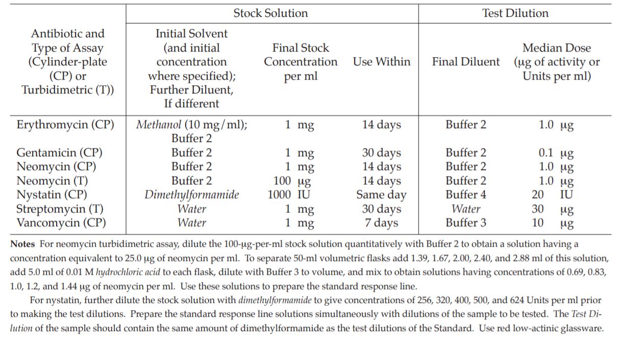
To prepare a stock solution, dissolve a quantity of the Reference Substance of a given antibiotic, accurately weighed, or the entire contents of a vial of Reference Substance, where appropriate, in the solvent specified in Table 1, and then dilute to the required concentration as indicated. Store in a refrigerator, and use within the period indicated. On the day of the assay, prepare from the stock solution five or more test dilutions, the successive solutions increasing stepwise in concentration, usually in the ratio of 1:1.25 for a cylinder-plate assay or smaller for a turbidimetric assay. Use the final diluent specified and a sequence such that the middle or median has the concentration designated.
Preparation of the Sample
From the information available for the preparation to be assayed (the “Unknown”), assign to it an assumed potency per unit weight or volume, and on this assumption prepare on the day of the assay a stock solution and test dilution as specified for each antibiotic but with the same final diluent as used for the Reference Substance. The assay with five levels of the Standard requires only one level of the Unknown at a concentration assumed equal to the median level of the Standard.
Organisms and Inoculum
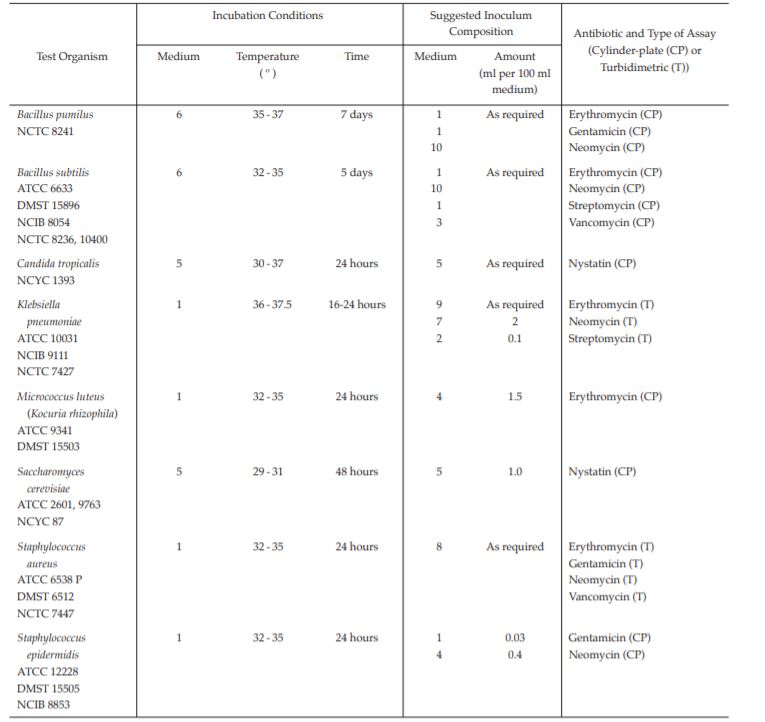
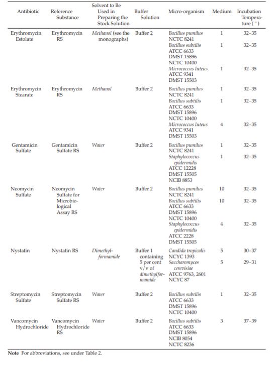
Test organisms The test organism for each antibiotic is listed in Table 2. The method of assay is given for each in Table 1. Maintain a culture on slants of the medium and under the incubation conditions specified in Tables 3 and 4 and transfer weekly to fresh slants. For Klebsiella pneumoniae use a noncapsulated culture.
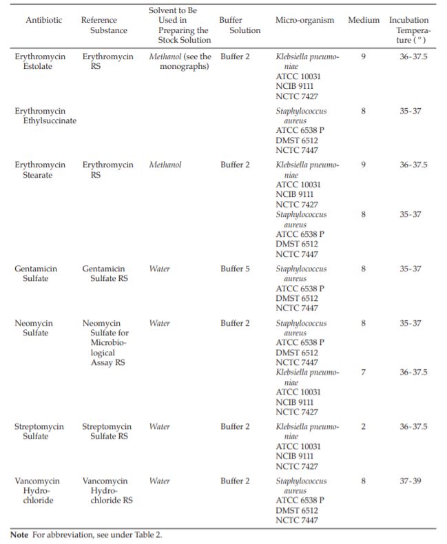
Table 1 Preparation of Stock Solutions and Test Dilutions of Reference Substances
 Preparation of inoculum
Bacillus subtilis; Bacillus pumilus Spore suspensions of the organisms to be used as inocula are prepared as follows. Grow the organism at 35º to 37º for 7 days1 on the surface of Medium 6. Using sterile water, wash off the growth, which consists mainly of spores. Heat the suspension at 70º for 30 minutes and dilute to give an appropriate concentration of spores, usually 10 × 106 to 100 × 106 per ml. The spore suspensions may be stored for long periods at a temperature not exceeding 4º. Alternatively, spore suspensions may be prepared by cultivating the organisms in Medium 8 at 26º for 4 to 6 days, then adding, aseptically, sufficient manganese sulfate to give a concentration of 0.001 g per litre and incubating for a further 48 hours. Examine the suspension microscopically to ensure that adequate spore formation has taken place (about 80 per cent) and centrifuge. Resuspend the sediment in sterile water to give a concentration of 10 × 106 to 100 × 106 spores per ml, and then heat to 70º for 30 minutes. Store the suspension at a temperature not exceeding 4º.
Klebsiella pneumoniae; Micrococcus luteus; Staphylococcus aureus; Staphylococcus epidermidis Grow the test organism on Medium 1 at 32º to 35º for 24 hours2 and adjust the opacity to one which has been shown to produce a satisfactory dose-response relationship in the turbidimetric assay, or to produce clearly defined zones of inhibition of convenient diameter in the diffusion assay, as appropriate.
Saccharomyces cerevisiae; Candida tropicalis Grow the test organism on Medium 5 at 29º to 31º for 48 hours3 . Wash off the growth with sterile saline TS. Dilute to a suitable opacity with the same solution. Determine by trial the quantity of stock suspension to be used as the inoculum, starting with the volume suggested in Table 2. The trial tests should be incubated for the times indicated in the section Turbidimetric method. Adjust the quantity of inoculum on a daily basis, if necessary, to obtain the optimum dose-response relationship from the amount of growth of the test organism in the assay tubes and the length of the time of incubation. At the completion of the incubation periods described in the section Turbidimetric method, tubes containing the median dose of the Standard should have absorbances of at least 0.3 absorbance unit.
For the cylinder-plate assay, determine by trial the proportions of stock suspension to be incorporated in the inoculum, starting with the volumes indicated in Table 2, that result in satisfactory demarcation of the zones of inhibition of about 14 to 16 mm in diameter and giving a reproducible dose relationship. Prepare the inoculum by adding a portion of stock suspension to a sufficient amount of agar medium that has been melted and cooled to 45º to 50º, and swirling to attain a homogeneous suspension.
Procedure
Assay designs The potency of an antibiotic is estimated by comparing the inhibition of growth of sensitive micro-organisms produced by known concentrations of the antibiotic to be examined and a reference substance.
1Except for Bacillus subtilis (incubate at 32º to 35º for 5 days).
2Except for Klebsiella pneumoniae (incubate at 36º to 37.5º for 16 to 24 hours).
3Except for Candida tropicalis (incubate at 30º to 37º for 24 hours).
Table 2 Preparation of Inoculum
ATCC --- American Type Culture Collection 10801 University Boulevard, Manassas VA 20110-2209, USA (http://www.atcc.org)
DMST --- Department of Medical Sciences, Ministry of Public Health, Nonthaburi 11000, Thailand
NCIB --- National Collection of Industrial Bacteria Torry Research Station, PO Box 31, 135 Abbey Road, Aberdeen AB9 8DG, Scotland (Note In 1983, the management of the collection of industrial bacteria under NCIB was transferred to the National Collection of Industrial and Marine Bacteria Ltd (NCIMB), 23 St Machar Drive, Aberdeen AB2 1RY, Great Britain.)
NCTC --- National Collection of Type Cultures Central Public Health Laboratory, Colindale Avenue, London NW9 5HT, Great Britain
NCYC --- National Collection of Yeast Cultures AFRC Food Research Institute, Colney Lane, Norwich NR4 7UA, Great Britain
The assay must be designed in a way that will permit examination of the validity of the mathematical model on which the potency equation is based. If a parallel-line model is chosen, the two log dose-response (or transformed response) lines of the preparation being examined and the reference preparation must be parallel; they must be linear over the range of doses used in the calculation. These conditions must be verified by validity tests for a given probability, usually P = 0.05. Other mathematical models, such as the slope ratio model, may be used provided that proof of validity is demonstrated (Appendix 9).
Unless otherwise stated in the monograph, the confidence limits (P = 0.95) of the assay for potency are not less than 95 per cent and not more than 105 per cent of the estimated potency.
Carry out the assay by the diffusion or turbidimetric method as listed in Tables 3 and 4.
Diffusion method (Cylinder-plate method) Liquefy a medium suitable for the conditions of the assay and inoculate it at a suitable temperature, for example, 48º to 50º for vegetative forms, with a known quantity of a suspension of micro-organisms sensitive to the antibiotic being examined, such that clearly defined zones of inhibition of suitable diameter are produced with the concentrations of the antibiotic used for the assay. Immediately pour into Petri dishes or large rectangular dishes a quantity of the inoculated medium to form a uniform layer 2 to 5 mm thick. Alternatively, the medium may consist of two layers, only the upper layer being inoculated.
Store the dishes so that no appreciable growth or death of the micro-organisms occurs before the dishes are used and so that the surface of the medium is dry at the time of use.
Using the solvent and the buffer solution indicated in Table 3, prepare solutions of the reference substance and of the antibiotic being examined having known concentrations and presumed to be of equal activity. Apply the solutions to the surface of the medium, for example, in sterile cylinders of porcelain, stainless steel or other suitable material, or in cavities prepared in the agar. The same volume of solution must be added to each cylinder or cavity. Alternatively, use sterile absorbent paper discs of suitable quality; impregnate the discs with the solutions of the reference substance or the solutions of the antibiotic to be examined and place on the surface of the agar.
In order to assess the validity of the assay, use not less than three doses of the reference substance and three doses of the antibiotic being examined having the same presumed activity as the doses of the reference substance. It is preferable to use a series of doses in geometric progression. In routine assays when the linearity of the system has been demonstrated over an adequate number of experiments using a three-point assay, a two-point assay may be sufficient, subject to agreement by the competent authority. However, in all cases of dispute, a three-point assay as described above must be applied.
Arrange the solutions on each Petri dish or on each rectangular dish according to a statistically suitable design, except for small Petri dishes that cannot accommodate more than six solutions, arrange the solutions of the antibiotic to be examined and the solutions of the reference substance in an alternate manner to avoid interaction of the more concentrated solutions.
Incubate at a suitable temperature for about 18 hours. A period of diffusion prior to incubation, usually 1 to 4 hours, at room temperature or at about 4º, as appropriate, may be used to minimize the effects of the variation in time between the application of the solutions and to improve the regression slope.
Measure the diameters with a precision of at least 0.1 mm or the areas of the circular inhibition zones with a corresponding precision and calculate the potency using appropriate statistical methods.
Use in each assay the number of replications per dose sufficient to ensure the required precision. The assay may be repeated and the results combined statistically to obtain the required precision and to ascertain whether the potency of the antibiotic to be examined is not less than the minimum required.
For the 1-level assay with a standard curve, prepare dilution representing five test levels of the Standard (S1 to S5) and a single test level of the Unknown U3 corresponding to S3 of the standard curve defined under Preparation of the Standard and Preparation of the Sample. For deriving the standard curve, fill alternate cylinders as each of three plates with the median test dilution (S3) of the Standard and each of the remaining nine cylinders with one of the other dilutions of the Standard. Repeat the process for the three dilutions of the Standard. For each Unknown, fill alternate cylinders on each three plates with the median test dilution of the Standard (S3), and the remaining nine cylinders with the corresponding test dilution of the Unknown (U3).
ESTIMATION OF POTENCY To establish a standard response line (standard curve), average the 36 inhibition zone diameters of the standard median concentration in all the four groups of plates. Use this value as the “corrected, median point” of the standard curve. Average also the nine inhibition zone diameters of the standard median concentration in each of the four groups of three plates. Correct the average diameter obtained for each concentration of the standard test solutions to the figure it would be if the average median concentration diameter for that group of three plates were the same as the corrected median point. Thus, if in correcting the figure for the highest concentration of the standard curve, the average of the 36 diameters of the median concentration, the corrected median point, is 16.5 mm, and if the average of the median concentration of the group of three plates containing the highest concentration of the standard test solution is 16.3 mm, the correction is +0.2 mm. If the average reading of the inhibition zone diameter of the highest concentration of
Table 3 Diffusion Method
the standard test solution of these same three plates is 16.9 mm, the corrected diameter is then 17.1 mm. Plot these corrected diameters, including the corrected median point, on a two-cycle semilog paper, using the concentrations of the antibiotic in micrograms or units per ml as the ordinate (logarithmic scale) and the diameters of the inhibition zones as the abscissa (arithmatic scale). Thus, the response line is drawn through points plotted for the highest and lowest zone diameters obtained by means of the following equations:
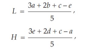
where L = calculated zone diameter for the lowest concentration of the standard response line,
H = calculated zone diameter for the highest concentration of the standard response line,
c = average zone diameter of 36 readings of the median point standard solution,
a, b, d, and e = corrected average values for the other standard solutions, lowest to highest concentration, respectively
To estimate the potency of the Unknown, average the zone diameters of the Standard and the zone diameters of the Unknown on the three plates used. If the average zone diameter of the Unknown is larger than that of the Standard, add the difference between them to the median concentration diameter of the standard response line. If the average zone diameter of the Unknown is lower than that of the Standard, subtract the difference between them from the median concentration diameter of the standard response line. From the response line, read the concentration corresponding to these corrected values of zone diameters. Multiply the concentration by the appropriate dilution factor to obtain the antibiotic content of the Unknown.
Turbidimetric method (Tube method) Inoculate a suitable medium with a suspension of the chosen microorganism having a sensitivity to the antibiotic being examined such that a sufficiently large inhibition of microbial growth occurs in the conditions of the test. Use a known quantity of the suspension chosen so as to obtain a readily measurable opacity after an incubation period of about 4 hours.
Use the inoculated medium immediately after its preparation.
Using the solvent and the buffer solution indicated in Table 4 prepare solutions of the reference substance and of the antibiotic being examined having known concentrations presumed to be of equal activity.
In order that the validity of the assay may be assessed, use not less than three doses of the reference substance and three doses of the antibiotic to be exam ined having the same presumed activity as the doses of the reference substance. It is preferable to use a series of doses in geometric progression. In order to obtain the required linearity, it may be necessary to select from a large number three consecutive doses, using corresponding doses for the reference substance and the antibiotic being examined.
Distribute an equal volume of each of the solutions into identical test-tubes and add to each tube an equal volume of inoculated medium (for example, 1 ml of the solution and 9 ml of the medium). Prepare at the same time two control tubes without antibiotic, both containing the inoculated medium and to one of which is added immediately 0.5 ml of formaldehyde solution. These tubes are used to set the optical apparatus used to measure the growth.
Place all the tubes, randomly distributed or in a Latin square or randomized block arrangement, in a water-bath or other suitable apparatus fitted with a means of bringing all the tubes rapidly to the appropriate incubation temperature and maintain them at that temperature for 3 to 4 hours, taking precautions to ensure uniformity of temperature and identical incubation time.
After incubation, stop the growth of the microorganisms by adding 0.5 ml of formaldehyde solution to each tube or by heat treatment and measure the opacity to three significant figures using suitable optical apparatus. Alternatively, use a method which allows the opacity of each tube to be measured after exactly the same period of incubation.
Calculate the potency using appropriate statistical methods (Appendix 9).
Linearity of the dose-response relationship, transformed or untransformed, is often obtained only over a very limited range. It is this range which must be used in calculating the activity and it must include at least three consecutive doses in order to permit linearity to be verified. In routine assays when the linearity of the system has been demonstrated over an adequate number of experiments using a three-point assay, a twopoint assay may be sufficient, subject to agreement by the competent authority. However, in all cases of dispute, a three-point assay must be applied.
Use in each assay the number of replications per dose sufficient to ensure the required precision. The assay may be repeated and the results combined statistically to obtain the required precision and to ascertain whether the potency of the antibiotic being examined is not less than the minimum required.
For the 1-level assay with a standard curve, prepare dilutions representing five test levels of the Standard (S1 to S5) and a single use level (U3) of each of up to 20 Unknowns corresponding to S3 of the Standard. Prepare also an extra S3 as a test of growth. Add 1 ml of each test dilution to three tubes and 1 ml of antibioticfree diluent to six tubes as controls. Distribute one complete set, including two tubes of controls to a tube rack, intermingling them at random. Add 9.0 ml of inoculum, incubate, add 0.5 ml of diluted formaldehyde
Table 4 Turbidimetric Method
solution (1 in 3), and complete the assay as directed above. Determine the exact duration of incubation by observation of growth in the reference concentration (median dose) of the dilutions of the Standard (S3).
ESTIMATION OF POTENCY To prepare the standard response line, plot the average transmittance or absorbance values for each concentration of the standard response line on a one-cycle semilog paper, with the transmittance or absorbance values on the arithmatic scale and the concentrations on the logarithmic scale. The response line is drawn through points plotted for the highest and lowest transmittance or absorbance values by means of the equations stated in Plate method.
To estimate the potency of the Unknown, average the transmittance or absorbance values of the Unknown and determine the antibiotic concentration from the standard response line. Multiply the concentration by the appropriate dilution factor to obtain the antibiotic content of the Unknown.